Home > Conditions Treated > Cerebrovascular Diseases > Arteriovenous Malformation (AVM)

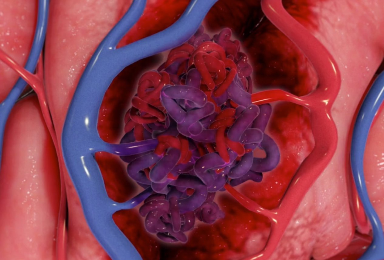
An arteriovenous malformation (or AVM) involves abnormal communication between the brain arteries and veins.

What is an arteriovenous malformation?

An arteriovenous malformation (or AVM) involves abnormal communication between the brain arteries and veins. Normally, high pressure blood travels to the brain through arteries. These arteries become progressively smaller and smaller. They terminate into tiny capillaries that supply brain tissue with blood. Low pressure blood then travels away from the brain through thin-walled veins.

In an AVM, there is direct communication between arteries and veins without the intervening brain tissue. This means that high pressure arterial blood passes directly into a thin walled vein. This may rupture and cause bleeding in the brain.

AVMs are thought to be congenital lesions (present from birth) and can rupture in childhood or adulthood. An AVM may also cause seizures or stroke-like symptoms or it may be discovered incidentally.

Risks Of Arteriovenous Malformation Rupturing

The risk of bleeding of a ruptured AVM is estimated to be approximately 3% per year. In the short-term the rupture risk is therefore small but in the long term the rupture risk adds up. In young and middle aged patients, the overall lifetime risk of rupture is substantial.

If an AVM ruptures, the risk of re-bleeding from the same AVM is approximately 6% per year for the following 2 years. Then gradually, over a period of a few years, the risk reduces to the baseline of approximately 3% per year.

Ruptured Arteriovenous Malformation Prognosis

A ruptured AVM usually causes a haemorrhage within the brain tissue. Depending on the size and location of the haemorrhage, approximately 10% of patients die from the haemorrhage. Up to a third of patients may be left with permanent disability. If a ruptured AVM causes a large haemorrhage, this requires urgent surgery to evacuate the haemorrhage and resect the AVM. This is usually performed by a cerebrovascular neurosurgeon with experience in AVM surgery.

Arteriovenous Malformation Treatment

When deciding on whether to treat an un-ruptured AVM, it is necessary to consider the estimated lifetime risk of rupture compared to the estimated risk of treatment.

The size, location, and venous drainage of an AVM are taken into account when determining the risk from surgical removal.

With some AVMs the estimated risk of surgical removal is significantly smaller than the lifetime risk of rupture and therefore surgical removal is a recommended option. Whereas other AVMs are too risky to resect surgically and it may be safer not to operate on them.

Micro-surgery is still considered the best treatment option when it’s performed safely. This is because it carries around 95% chance of instant cure. In some cases, microsurgery is too risky and other treatment options can be considered. These include stereotactic radiosurgery (SRS) or embolisation.

Stereotactic radiosurgery

Stereotactic radiosurgery involves using computer guidance to concentrate beams of radiation to the AVM. This slowly causes abnormal vessels to close off. The success rate from SRS is generally lower than microsurgery.  It can also take up to 3 years before the AVM is cured and during this period the AVM can still rupture.

Embolisation

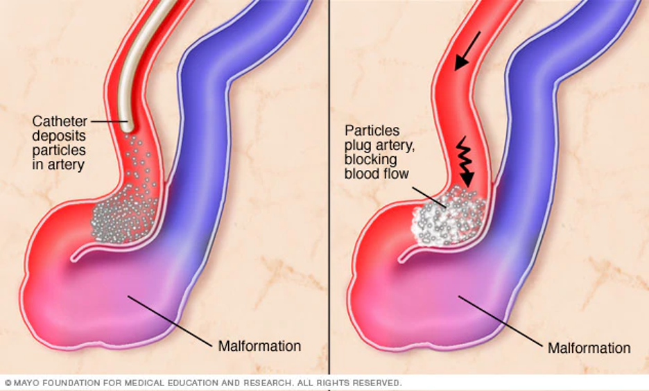
Embolisation involves injecting liquid glue or particles into the AVM. This method inserts microcatheters through an artery in the groin. The success rate of embolisation alone in curing an AVM is often poor.  Embolisation can still be used in combination with microsurgery or SRS.

The decision to treat an un-ruptured AVM must be taken by the patient. This follows extensive discussions and guidance from an experienced multi-disciplinary team consisting of a cerebrovascular neurosurgeon, an interventional neuroradiologist and a radiation therapist.

Click here to return to the cerebrovascular diseases page.